By utilizing our innovative and proprietary lipid conjugation technology, LipoD293â„¢ (Ver. II) is specially designed and formulated by adding proprietary enhancers for transfecting HEK293 cells and other mammalian cells (Figure 1). As a 2nd generation liposome based DNA transfection reagent, LipoD293â„¢ (Ver. II) offers extremely high transfection efficiencies for HEK293 related cells as well as many mammalian cells with less cytotoxicity. LipoD293â„¢ reagent (Ver. II), upgraded from its previous version with refined chemistry, is 3~4 times more efficient in gene delivery. LipoD293â„¢ reagent (Ver. II), 1.0 ml, is sufficient for ~666 transfections in 24 well plates or ~333 transfections in 6 well plates.
Figure 1. A Cartoon Showing LipoD293™ (Ver. II) Enhanced Gene Delivery
Features
- Top choice for hard-to-transfect cells
- Exceptional high titers of virus production
- Equally good for very long DNAs (up to 180 kb)
- Equally good for suspension 293 cells (e.g., 293F, 293H, etc)
- High levels of recombinant protein production
- Presence of serum and antibiotics enhances efficiency on 293 cells
- Exceptional efficiency for both single DNA transfection and multi DNAs co-transfection
- Very affordable
Comparisons of Transfection Efficiency of LipoD293â„¢ DNA In Vitro Transfection Reagent (Ver. II) with Brand Name Products
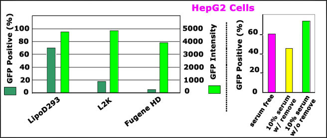
Comparison of transfection efficiency of LipoD293â„¢ reagent (Ver. II) vs. lipofectamine 2000 (L2K) and Fugene HD on HepG2 cells.
Right Panel: Comparison of transfection efficiency of LipoD293 (Ver. II) with Lipofecatmine 2000 (L2K), and Fugene HD on HepG2 cells. GFP DNA (pEGFP-N3) was transfected with different transfection reagents per manufacturer's protocols to HepG2 cell (cultured on Collagen pretreated dishes). GFP positive cell (%) and fluorescence intensity were detected by passing through FACS 48 hours post transfection
Left Panel: presence of serum and antibiotics enhances LipoD293 (Ver. II) efficiency on HepG2 cells. HepG2 cell (grown on collagen treated dishes) was transfected with three different conditions-------serum and antibiotics free, presence of 10% serum and antibiotics followed by removal 5 hours post transfection and presence of 10% serum and antibiotics without removal 5 hours post transfection.
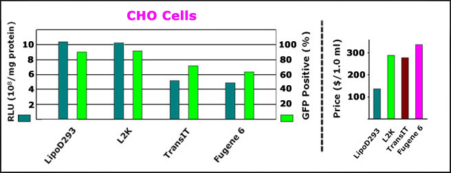
Comparison of transfection efficiency of LipoD293â„¢ reagent (Ver. II) vs. lipofectamine 2000 (L2K), TransIT and Fugene 6 on CHO cells.
Right Panel:Â Comparison of transfection efficiency of LipoD293 (Ver. II) with Lipofecatmine 2000 (L2K), TransIT and Fugene 6 on CHO cells. DNAs encoding Renilla luciferase (phRL-CMV) and GFP (pEGFP-N3) were transfected with different DNA transfection reagent per manufacturer's protocols. Renilla luciferase activity and GFP fluorescence were detected with Renilla Assay System and a Nikon Eclipse fluorescent microcopy respectively 24 hours post transfection.
Left Panel:Â Comparison of price ($/1.0 ml vial) of LipoD293 versus those of Lipofecatmine 2000 (L2K), TransIT and Fugene 6. All the prices were collected from the manufacturers' websites.
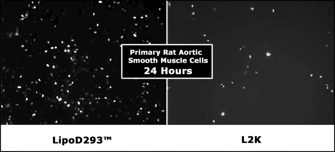
A comparison of transfection efficiency of LipoD293™ reagent with lipofectamine 2000 (L2K) on a hard-to-transfect cell, primary rat aortic smooth muscle cells. The rat aortic smooth muscle cells were prepared and transfected with pEGFP-N3 by LipoD293™ reagent (left panel) and Lipofecatmine 2000 (L2K, right panel) respectively per manufacturers' protocols. The transfection efficiency was evaluated by detecting GFP fluorescence with a Nikon Eclipse 2000 microscopy 24 hours post transfection. The above pictures were kindly provided by Dr. Nickolai Dulin of Section of Pulmonary and Critical Care, University of Chicago.
A comparison of transfection efficiency of LipoD293™ reagent with Fugene HD on hard-to-transfect cell, LNCap cells. The LNCap cells were grown as ATCC recommended procedures and co-transfected with pBabe-hygro-SSeCKs (1.5ug) and pEGFP-N3 (0.5ug) per well (6 well plate) LipoD293™ reagent (left panel) and Fugene HD (right panel) respectively per manufacturers' protocols. The transfection efficiency was evaluated by detecting GFP fluorescence with a Nikon Eclipse 2000 microscopy 24 hours post transfection. The above pictures were kindly provided by Dr. Lyn Gao of Roswell Park Cancer Institute.
Two examples showing exceptional efficiency of LipoD293™ reagent on hard-to-transfect cells like HepG2 and SaoS-2 cells. HepG2 and SaoS-2 cells in 95% confluency were transfected with pEGFP-N3 and pSV-?-galactosidase DNAs respectively in presence of serum/antibiotics. The efficiency was checked 48 hours post transfection by Zeiss 510 Confocal Microscopy and ?-galactosidase staining kit respectively.

A comparison of transfection efficiency of LipoD293™ reagent with 293fectin on HEK293 cells. HEK293 cells transfected with pEGFP-C1 plasmid using LipoD293™ In Vitro DNA Transfection Reagent (Ver. II) (upper panel) and the most popular brand product 293fectin™ of Invitrogen (lower panel). The cells were visualized by Nikon Eclipse Fluorescence microscope with DIC phase imaging (left panel) and FITC imaging (right panel) 24 hours post-transfection.
A comparison of LipoD293™ reagent vs. 293fectin, Xfect and Fugene 6 transfection reagents on protein production with suspension 293F cells. 30 mL of 293F cell cultured in standard culture medium was transfected with pEGFP-6xHis plasmid using LipoD293™ In Vitro DNA Transfection Reagent (Ver. II) (20 ug plasmid DNA), 293Fectin (30 ug plasmid DNA), Xfect (30 ug plasmid DNA) and Fugene 6 (30 ug plasmid DNA) per manufacturers' standard transfection protocols. GFP fluorescence was visualized 48 hours post transfection (left panel) with A for LipoD293, B for 293fectin, C for Xfect and D for Fugene 6. The 6xHis tagged GFP protein was then purified via Ni-NTA affinity column. 5 ul of 1st elution fraction was resolved on SDS-PAGE followed by Coomassie Brilliant Blue staining (right upper panel E) with the lane 1 for LipoD293, lane 2 for protein marker, lane 3 for Xfect, lane 4 for 293fectin and lane 5 for Fugene 6. The protein yield was quantified via spectrometer (right lower panel F).

A comparison of LipoD293â„¢ (Ver. II) and Lipofecatmine LTX (LTX) on generation of Lentivirus (LV). Three cDNAs were co-transfected with LipoD293â„¢ (Ver. II) and Lipofectamine LTX (LTX) into 293FT cells. A GFP vector, pHR-SIN-cppt-CMVEWP, was used to determine titer of LV. 1x10⁵ 293F cells per well was plated in to a 24 well plate followed by addition of different of amounts of the vector supernatant, 1 microliter (upper panel) and 10 microliters (lower panel) respectively. 5 days later, the cells was passed with FACS. The numbers at the upper right corner indicate the percentage of transduced cells. The titers of LV generated with LipoD293â„¢ and L2K were quantified to be 8x10^6 and 3x10^6 tu/ml respectively